


31st January 2025
Almost 80% of “refugees” go on vacation to the country they claim to have fled from …
Quote


@Rothmus
31st January 2025
So, not “refugees”

31st January 2025
Quote
@Rothmus
31st January 2025
So, not “refugees”
Amanpour and Company475K subscribers
Jan 28, 2025 #amanpourpbsFor Palestinians returning to their homes in Northern Gaza, and for families in Israel reuniting with their loved ones, conversation now begins to turn to rebuilding, processing, and healing. Renowned physician, author and self-help guru Dr. Gabor Maté, a Holocaust survivor, has an intimate understanding of the process. He joins Michel Martin to discuss how his own life experience has informed his perspective on trauma in Israel and the Palestinian territories. Originally aired on January 28, 2025

FeaturedNeuroscienceVisual Neuroscience
·January 27, 2025
Summary: New research highlights a functional hierarchy in the brain’s processing of space and time. In posterior areas, like the occipital cortex, space and time are tightly linked and processed by the same neurons.
In anterior regions, such as the frontal cortex, space and time are processed independently, with distinct neural populations forming “time maps” for specific durations. Intermediate regions, like the parietal cortex, display mixed processing mechanisms, bridging spatial and temporal integration.
This study offers fresh insights into how the brain integrates two fundamental dimensions of human experience and reveals the unique coding strategies across cortical regions.
Key Facts:
Source: SISSA
Imagine a swarm of fireflies, flickering lights on and off in the nighttime space. How does the human brain process and integrate information about duration and spatial position enabling this vision?
This was the question addressed by Valeria Centanino, Gianfranco Fortunato, and Domenica Bueti from SISSA’s Cognitive Neuroscience group in their research published in Nature Communications.
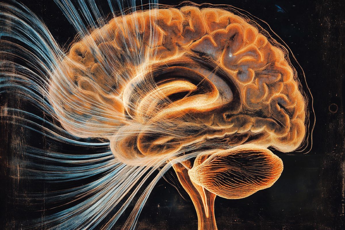
The study highlights the existence of a functional hierarchy in the human cerebral cortex, where posterior areas—those that first receive visual information— process space and time together.
In contrast, in parietal areas and even more so in frontal areas, engaged later in information processing, space and time gradually separate. Moreover, the way time is encoded differs across these brain regions.
In the occipital areas, where space and time are processed together, time is encoded in the activity of the same neural population, which becomes proportionally more active the longer the duration.
In parietal and frontal areas, instead, where the link between space and time becomes progressively weaker and eventually disappears, time is encoded by distinct neural populations, each of them responding selectively to specific durations.
In the parietal areas, in an intermediate position of this hierarchy, there is a coexistence of duration coding mechanisms and time and space are either processed toghether or independently.
The Experiment: Visual Stimuli in Different Screen Positions
“How does the human brain process visual information changing in both spatial position and duration?”
This was the question driving the research.
“We wanted to understand whether space and time are processed independently or not,” the authors of the study explain. To simulate what might occur in reality, they asked young healthy participants to judge the duration of a visual stimulus presented at different positions on a screen for varying amounts of time.
“During this task, neural responses were recorded using high-resolution spatial magnetic resonance imaging (fMRI).”
Different Brain Areas, Different Coding Strategies. Posterior Cortex: Space and Time Processed Together
“What we discovered is that the link between space and time in the human brain is not fixed but depends on the brain areas involved. In the posterior part of our brain, particularly in the occipital visual cortex, the connection is strong because space and time are processed by the same neurons.
“This region, specialized in receiving visual inputs,” explain Centanino, Fortunato, and Bueti, “responded to both the position and duration of the stimulus: the longer the viewing time, the greater the brain activity in these neuron populations.”
Anterior Cortex: Separate Neurons for Space and Time
In the anterior regions, however, this connection disappears. In the frontal premotor areas, involved in movement preparation, time is processed independently of the position of the visual stimulus.
Distinct populations of neurons process space and time. Furthermore, the authors add, “duration is encoded differently compared to the posterior areas.
Here, distinct neural populations preferentially respond to specific durations, and neural populations that prefer similar durations are contigous in the cortical surface, forming what we might call ‘time maps.’”
Intermediate Cortex: A Variety of Mechanisms
In an intermediate region of this cortical hierachy, specifically the parietal cortex, which is known for integrating various information sources and is functionally situated between the occipital and frontal premotor cortex, the relationship between space and time is multifaceted.
The three researchers explain: “Some neuronal populations responded to both the position and the duration of the stimulus, while others responded only to one of these dimensions.
“The response to time was, in some cases, monotonic like in the occipital cortex, while in others it showed selectivity for specific durations, similar to the anterior areas.”
A Functional Hierarchy in Time Processing
“This study,” conclude Centanino, Fortunato, and Bueti, “advances our understanding of how space and time, two fundamental aspects of our experience of the world, are processed and integrated in the human brain.
“Moreover, it sheds light on the presence of a functional hierarchy in time processing.
“The existence of multiple response profiles to stimulus duration, along with their specific relationship to spatial processing, suggests that different brain areas contribute distinctly to the processing and perception of time.”
Author: Donato Ramani
Source: SISSA
Contact: Donato Ramani – SISSA
Image: The image is credited to Neuroscience News
Original Research: Open access.
“The neural link between stimulus duration and spatial location in the human visual hierarchy” by Valeria Centanino et al. Nature Communications

The offices where Washington D.C.’s bureaucrats work were largely empty on Monday, January 20, 2025, as Donald Trump was inaugurated as U.S. President for the second time.
But they weren’t empty for the inauguration. Because that Monday coincided with the Martin Luther King Jr. Day federal holiday, most U.S. government employees had a paid day off work.
But then, many of the federal government’s buildings continued to sit empty on Tuesday, January 21, and again on Wednesday, Thursday, and Friday.
During that time, the bureaucrats discovered they greatly liked working from home. So much so that even after President Biden declared the pandemic a national emergency on April 11, 2023, they kept doing it. It’s become quite a popular perk among the ranks of the U.S. government’s civilian employees.
Senator Joni Ernst issued a report on the cost to taxpayers of the bureaucrats’ latest addition to their benefits. Here are some excerpts of her report with its major takeaways:
Just three percent of the federal workforce teleworked on a daily basis before the COVID-19 pandemic. Today, the temporary pandemic-era practice is a presumed public employee perk. Six percent report in-person on a full-time basis while nearly a third of the government workforce is entirely remote.
Ernst included several anecdotes from whistleblowers to accompany her findings. Here’s one for the U.S. Department of Agriculture:
A whistleblower who is a current supervisor within the U.S. Department of Agriculture (USDA) informs me “the vast majority of USDA employees are not working in person. On the occasions I have gone to USDA headquarters in Washington, D.C., it resembles a ghost town. Hallways are mostly empty, and offices are unoccupied.” This whistleblower says, “remote work and telework employees are often unreachable and do not respond to simple email questions for hours. This leads to inefficiency in completing tasks in a timely manner and to delays in clearing documents and reports due to the inability to reach colleagues.”
Here’s another from a government employee who requested anonymity because they are among the very few showing up at the office:
A federal employee who wished to remain anonymous says “he is one of few who reports to the Washington, D.C., office, and contractors have commented to him about the whereabouts of agency employees.” He observes, “it’s all empty around me. I’m the only person within three rows where I sit. It doesn’t look good.”
Here’s a final note about the Department of Veterans Affairs, highlighting a bureaucrat behaving poorly:
When a Department of Veterans Affairs (VA) manager posted a picture of himself “working” from a bubble bath on social media, his frustrated coworkers became whistleblowers. One of which stated, “If you think that this is not a big deal then what is a big deal? Is it a big deal when a veteran dies?” That’s right, the employees of the agency turned in their own manager for abusing telework.
To Americans who work in the real world, it’s obvious the bureaucrats’ continuing absence from the office cannot be sustained. It’s no accident that one of President Trump’s first-day executive orders was directed at teleworking bureaucrats. On January 23, the government’s Office of Personnel Management ordered all federal agencies to comply with that executive order within 30 days.
FacebookTwitterEmailFlipboardMastodonLinkedInShare

Craig Eyermann is a Research Fellow at the Independent Institute. He is also the creator of MyGovCost.org: Government Cost Calculator. He received his M.S. in mechanical engineering from New Mexico State University and M.B.A. from the University of Phoenix, having received a B.S. in both mechanical and aerospace engineering from the Missouri University of Science and Technology.

A group of scientists who are experts at spotting fraudulent research outline five essential steps to combat industrialized scientific misconduct
‘Stamp Out Paper Mills …. Science Sleuths on how to fight.

Subscribe
The Barbary slave trade in which Europeans were abducted and sold into slavery from as far afield as Iceland and Ireland

| Trump Takes Aim at Public Health Last week Donald Trump was inaugurated for his second term. And this one is shaping up to be just as disruptive as his first for the health and research world. On January 20 Trump signed an executive order announcing he will withdraw the U.S. from the World Health Organization—the premier source for handling global disease outbreaks and promoting health care access. He tried to withdraw during his first term, but the process requires a year’s notice, so Biden blocked it. This time, the withdrawal is likely to go through. The WHO has faced its share of criticism for how it handled the COVID pandemic and the fact that it lacks powers of enforcement. But the U.S. withdrawal is bad news for the organization and its nearly 200 member countries, who rely on the U.S. for both funding and public health expertise. And it’s also bad for the U.S. because we will no longer have a seat at the table for important global health conversations—a gap China will likely fill. Also last week, the Trump administration froze all external communications from the Department of Health and Human Services, including the FDA and the CDC, at least through February 1. This means the CDC can’t release its weekly reports on disease outbreaks, for example, and the NIH—the country’s leading source of biomedical research funding—had to abruptly cancel meetings and grant reviews. It’s unclear how long the communications freeze will last, but it’s likely already impacting the government’s ability to get out timely health information, including about bird flu. Trump also signed a number of other executive orders defining biological sex as male or female, rolling back protections for transgender people, and reversing some of Biden’s attempts to lower drug prices for people on Medicare and Medicaid. And later this week, a confirmation hearing will be held for Robert F. Kennedy, Jr., Trump’s controversial pick for head of Health and Human Services. —Tanya Lewis |
January 23, 2025

AI detects lung diseases from the tiniest of ultrasound details
Generated by DALL-E
View 3 Images View gallery – 3 images
A breakthrough new AI model is able to detect the presence of different lung diseases from ultrasound videos, with 96.57% accuracy, and it is even able to distinguish whether the abnormalities are due to pneumonia, COVID-19 or other conditions.
The model, developed by researchers at Australia’s Charles Darwin University (CDU), United International University and the Australian Catholic University (ACU), can identify specific patterns of different lung disease, outperforming previous AI tools that have been tested on the same ultrasound datasets.
“The model also uses AI techniques to show radiologists why it made certain decisions, making it easier for them to trust and understand the results,” said study co-author Niusha Shafiabady, a professor at CDU. “This model helps doctors diagnose lung diseases quickly and accurately, supports their decision-making, saves time, and serves as a valuable training tool.”
The team combined two kinds of AI models, highlighting how adaptable the technology is for diagnostic needs. One, known as a convolutional neural network (CNN), seeks out patterns in images or frames, focusing on the tiniest pixel-based changes that the human eye can miss when examining scans. Then, a long short-term memory (LSTM) model uses this information and puts it into a broader context, analyzing the CNN’s data over time while ‘forgetting’ irrelevant data.
With their powers combined, the novel hybrid model known as TD-CNNLSTM-LungNet is able to pick up abnormalities incredibly well – and then explain what the issue is. What’s more, it can determine if the scans show evidence of pneumonia, COVID-19, other lung diseases or if the lungs are normal. With a high ‘recall’ rate of 96.51%, this essentially means there are very few false negatives identified by the AI – which is important in treating time-critical lung conditions.
Using ultrasound videos of existing datasets, the model surpassed existing AI diagnostic tools, which currently score around 90-92%.
For example, just a year ago the Food and Drug Administration (FDA) approved the use of the DermaSensor device, the first smartphone AI-powered device that was shown to be able to detect around 200 different kinds of skin cancers. (Incidentally, I had a basal cell carcinoma identified through a similar smartphone camera device and AI software.) While the tools are not intended to replace medical professionals – my skin-cancer specialist correctly identified the cancerous spot on my back before using the AI camera device – they are set to become the most beneficial and affordable technology we’ve ever seen in medicine.
This new lung disease AI model gives us a clue as to what is yet to come, with it able to correctly identify nuances that differentiate, for example COVID-19 from pneumonia. As the researchers noted, both of those conditions appeared similar to the human eye, but had distinct patterns that enabled the AI model to spot the difference. It would then produce a report as to why it came to its conclusion for each scan.
“The explainability of the proposed model aims to increase the reliability of this approach,” said Shafiabady. “The system shows doctors why it made certain decisions using visuals like heat maps. This interpretation technique will aid a radiologist in localizing the focus area and improve clinical transparency substantially.”
In 2024, huge strides were made by Google in the area of medical diagnostics and AI. Similarly, the technology is being developed for healthcare to assist with everything from surgery to drug discovery. It’s already shown its potential for use in detecting brain tumors and other cancers.
Remix News & Views (@RMXnews) posted at 0:34 pm on Tue, Jan 28, 2025:
NEW:

Germany’s federal health minister Karl Lauterbach claims that as many as 30 percent of asylum seekers entering Germany are “mentally ill” and warned that without therapy, “nobody can deny they represent a threat.”
Instead of refusing entry to those deemed dangerous to others, the Social Democrat wants Germany to ramp up its mental health services to treat people more quickly. “It can’t go on like this,” he adds.

https://t.co/ypOYgaaIAq
(https://x.com/RMXnews/status/1884218588178821148?t=_PVRXYHOdCRYpDX6fijqMg&s=03)

