
=================================================
Unveiling Opus Dei: Irishwoman from FT investigation speaks out
Updated / Saturday, 30 Mar 2024 15:42
Ann Marie Allen was 15 when she joined training course at an Opus Dei-run catering school. By 16 she had become an ‘assistant numerary’ within the organisation. During her years with Opus Dei, she worked from early morning until late evening in places like the organisation’s students’ residence in Galway, cooking and serving meals, doing laundry and cleaning rooms.
While not working she lived in an Opus Dei centre. There, she says, she was pressured to attend mass, deprive herself at meals, sleep on the floor one night a week, and tie a ‘cilice’ – “a barbed wire with the sharp bits on the inside” – around her leg for two hours daily.
She says she was isolated from her family, that her post was monitored, and her phone calls listened in to.
Eventually, with the help of her father, she says she “escaped” and rebuilt her life. Now, decades later, she has become a key part of a global effort by former assistant numeraries to unveil what happened within Opus Dei, and demand reparation.
Ms Allen’s story was central to a major investigation published by the Financial Times two weeks ago. In her first interview with Irish broadcast media, she told Katie Hannon on ‘Upfront: The Podcast’ further remarkable details about what she experienced within the organisation.
Listen: Katie Hannon speaks to Ann Marie Allen on Upfront: The Podcast
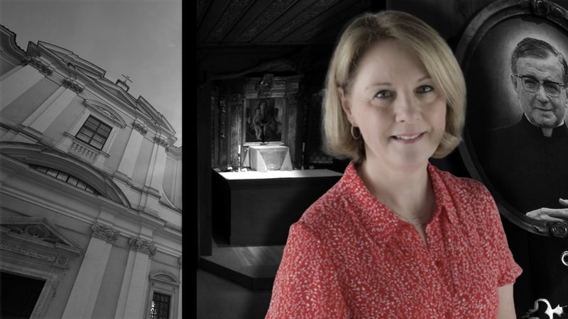
Opus Dei is a conservative Catholic institution which was founded by Saint Josemaría Escrivá in 1928. It now has a presence in over 60 countries. It consists of lay members and clerical members.
Upfront asked Opus Dei to respond to the details of Ann Marie’s story. In a statement, it said “we reject the accusation of exploitation,” adding “we are very sorry and deeply regret that Anne Marie Allen was hurt by her time in Opus Dei.”
Ms Allen says she was not paid during the seven years she was working in Opus Dei in the late 70s and early 80s. She wants the organisation to abolish the assistant numerary grade itself.
The Opus Dei statement says “Assistant numeraries are women in Opus Dei who, like all the other members, aim to love God and others through their work and daily life. In their case, their chosen work is caring for the people and centres in the family setting of Opus Dei.”
“This work is paid in accordance with the employment legislation of the countries in which they live.”
Ms Allen said she initially came into contact with Opus Dei when she looked at starting a course in a residential catering training school which was run by the organisation.
“We went for the interview. I remember both of my parents were taken away separately and I was taken away separately to be interviewed. We got the royal treatment… there was tea and coffee in beautiful surroundings,” she told Katie Hannon.
Love-bombing
Having started the course, she found the college lacked the structure typical of conventional educational institutions.
“There were no textbooks, there was no timetable. The classes were ad hoc. There were some outside teachers who came in and did some classes with us, but I now realise that they were trying to recruit them as well,” Ms Allen said.
“We got up every morning, we had to go to Mass. Even though they had said we had a choice, there was pressure put on us if we did not go to Mass. Then we had our breakfast very quickly and we got into a green bus. We drove into Salthill, and arrived there about 8.30am, and we worked until 8pm, seven days a week.”

In Salthill, she says she worked in an Opus Dei student residence and conference centre.
“I was sent there maybe a week after I started in the college… We cooked all of the meals. We did house-keeping and waitressed, and we did the laundry.”
Ms Allen says she was ‘love-bombed,’ by more senior members of Opus Dei, and told she had a vocation to be an assistant numerary. Once she accepted the vocation, she says the love-bombing stopped and she was pressured to comply with conversative religious orthodoxies and behaviours.
She says she was told “of you don’t follow your vocation, you won’t have happy relationships… you’ll go to hell.”
“It was very gradual. The manipulation…” she said. “By the time you get to join Opus Dei, you’re frightened and you’re completely isolated.”
Mortification
She said members used the cilice on their legs for two hours a day.
“I was always extremely uncomfortable about it. I was 16 years of age, and it was just given to me and said this is what you do. I had never heard of it; I’d never seen anything like this. And you did it.”
“Then the discipline was kind of a small whip that you had to whip yourself on a Saturday as a mortification,” she added.
“It wasn’t the worst part, believe it or not,” she said, before explaining ‘fraternal corrections’ were issued, a form of verbal admonishment.
“[It] was behaviour control… And you had to take it. At the end of it, you’d have to say, ‘oh, thank you very much for saying that to me.’ You couldn’t challenge anyone. That was constant. That was every day.”
“We were told at the end of a shower to take a cold water blast. You had to deprive yourself of something for every meal. You had to sleep on the floor one night a week without a mattress. You had to sleep one night in the bed without a pillow.”
“My contact with my family and with any friends was closely monitored,” Ms Allen told Katie Hannon.
“All of my letters that I wrote out, were read. You left the envelope open, and you handed it in, then anything they got back – or that you got back – was opened. You may or you may not get those letters… if they felt they weren’t suitable,” she said.
Ms Allen said she felt the purpose was to force conformity and servitude.
“Suppressing sexuality, suppressing your own values, your own opinions, it was constant. It was like Catholicism on cocaine.”
Leaving Opus Dei
After several years, Ms Allen’s father became concerned about her treatment, having read media reports about Opus Dei, and he tried to get her to leave.
When Ms Allen sought to visit home, she says Opus Dei insisted that her father write a letter “saying that he wouldn’t keep me.”
“Whenever my father was on the phone, they would sometimes be standing around me praying. If somebody came to see me, they’d be in the oratory praying.”
She was accompanied by more senior members of the organisation on the visit home.
“Dad said, ‘she’s staying, she’s going nowhere.’ And they said, ‘but you wrote a letter to say you wouldn’t keep her.’ And he said, what I wrote was, ‘I won’t try to keep her,’ but I am keeping her.”
After several difficult years, Ms Allen went back into education and studied at night to obtain her leaving certificate, and eventually a degree and masters.
She became a senior prison officer, retiring from the Irish Prison Service as part of the management team in some of Ireland’s best-known prisons.
In more recent years, she says she has connected with other former members of Opus Dei, after being introduced to one by a prison chaplin.
“She put me in contact with an ex-numerary. We met for lunch, and I was flabbergasted at the amount of people that had left. I found speaking to her was hugely therapeutic. I found it very, very healing. I went on my own journey of trying to find former members,” Ms Allen said.
Ms Allen has since joined 43 other former assistant numeraries – most from South America – who have made a complaint to the Vatican about their treatment from Opus Dei.
“All of the people that worked, that were unpaid, they need to be paid their wages, they need to get compensation, they need to have their pension entitlements restored and they need to be reimbursed for all of the counselling that they had to get from their treatment,” she told Katie Hannon.
Listen to Ann Marie Allen speaking to Katie Hannon on Upfront: The Podcast here, on Apple Podcasts and on Spotify.
In response to the allegations raised in this podcast, Opus Dei issued the following statement to RTÉ.
We are very sorry and deeply regret that Anne Marie Allen was hurt by her time in Opus Dei. She got in touch with us some months back and we replied and invited her to get in touch again. We have also approached her through an independent person to suggest we meet but so far she has not taken up the offer.
We hope she will do so in the future.
Anne Marie describes two different periods. The first was when she was a student at Ballabbert School. The second, when she chose a vocation within the Catholic Church.
With regard to her experience in the late 70s, we reject the accusation of exploitation. Ballabbert School was a non-profit socio-educational initiative supervised by the competent authorities. At that time the school leaving age in Ireland was 15 and many girls left school and went directly into employment with no qualifications which limited their opportunities to progress in life. Schools like Ballabbert provided an opportunity to acquire an internationally recognised qualification rather than going directly into employment. In the case of Ballabbert the girls took the National Council for Home Economics Education (later City and Guilds) exams.
Assistant numeraries are women in Opus Dei who, like all the other members, aim to love God and others through their work and daily life. In their case, their chosen work is caring for the people and centres in the family setting of Opus Dei. This work is paid in accordance with the employment legislation of the countries in which they live.
The vocation of assistant numerary is being followed by thousands of women around the world with freedom, love and commitment, and has the same dignity as any other life choice. In fact, many women who joyfully live out this vocational call made a public plea a few months ago for their free and conscious choice to be respected and not demeaned.
In cases in the past where there may have been irregularities in social security contributions, or bad experiences within the organisation, Opus Dei recognises that these things can have happened, but needs the people concerned to make a formal complaint. They can do so here.
For more information: https://opusdei.org/en-ie/article/q-a-about-the-financial-times-article/


 Image source: Wikipedia
 Illustration: Aïda Amer/Axios. Photo: Peng Bin/VCG via Getty Images